Advancing Pediatric Neurosurgical Care Through Education and Innovation

Assistant Professor of Neurological Surgery and Neurosurgical Direction of the Montefiore Einstein Craniofacial Center, expanded his research team comprising medical students from the Albert Einstein College of Medicine, residents, fellows, and scientists, conducting innovative research using novel revolutionary technologies to enhance pediatric neurosurgical care while mentoring and educating the next generation of compassionate healthcare leaders. State-of-the-art 3D photography can monitor changes in head structures and cranial volume over time with meticulous detail, AI may revolutionize the evaluation of brain scans, and Magnetic Resonance Elastography (MRE) can unlock hidden properties of the brain previously unrecognized with standard MRI techniques. These new technologies may help better detect and identify neurosurgical conditions, predict clinical outcomes, improve efficiency, and provide quantitative measurements for faster and more accurate diagnoses to support clinical decision-making, improved outcomes and patient safety.
Dr. Kobets and his team published the results of their study on 3D imaging in craniofacial surgery this past year in the Journal of Neurosurgery: Pediatrics. Using a novel 3D mobile application (MirrorMe3 platform), the authors evaluated the surgical outcomes of two 20-month-old patients with disfiguring right parietal cephalohematomas who underwent skull recontouring procedures. The application's ability to generate accurate models of the skull using photographs, capture the severity of a calcified cephalohematoma, and quantify the changes in the contour of the skull before and after surgery (which can be viewed in real-time on a mobile device and computer) were shown, providing an efficient method for monitoring patients with simple topographic scans.
We are among the first in the nation using a mobile app to better understand and quantify dimensional and volumetric cranial changes before, during and after surgery for children who have cranial deformities, improving on the subjectivity of interpreting volume and dimension from 2D pictures to more accurate and objective 3D images. Our proof-of-concept work can ultimately revolutionize the standard of care for cranial deformities in children and further inform and guide parents and children regarding the outcomes of surgery and how the corrections progress over time. We are also currently conducting research to assess the utility of this novel 3D mobile application for monitoring patients with other skull shape pathologies such as craniosynostosis and positional plagiocephaly who are treated with helmet therapy.
Dr. Kobets and his research team are also conducting an observational study integrating AI into reading brain magnetic resonance imaging (MRI) scans of premature infants with intraventricular hemorrhage (brain bleeding) to better understand which treatments (and the timing of these treatments) will result in the best long-term functional outcomes to optimize treatments for future patients. Currently, measures of the brain and fluid spaces are largely qualitative, predominantly looking at ventricular volume rather than the complete picture of the intracranial compartment. We are developing novel brain segmentation technologies using AI and atlas-based semiautomated segmentation of brain bleeding and brain volumes which can allow a numerical volume to be calculated to quantify the measurement of the brain and fluid spaces and to see the changes in these spaces before and after treatment. This may revolutionize the evaluation of brain scans in the future and one day be integrated into the standard of care for these patients.
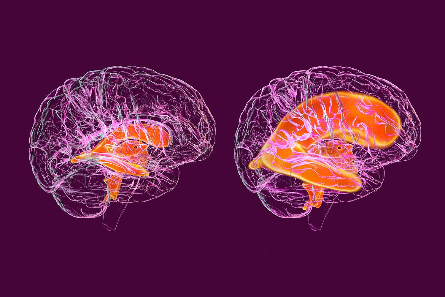
Our neurosurgeons and researchers are also using a new type of MRI, called Magnetic Resonance Elastography (MRE), to unlock hidden properties of the brain, previously unrecognized with standard MRI techniques, to gain an unprecedented understanding of the relationship between fluid spaces and the structural integrity and stiffness of brain tissue. We are one of only a few centers in the nation utilizing this technology to evaluate the brain. We published our research using MRE to identify the role that brain compliance plays in the progression and response to therapy (and failure thereof) in pediatric hydrocephalus and to elucidate the previously unappreciated consequences of shunt failures.
By combining MRI with special vibrations, or mechanical shear waves, this technique can further elucidate underlying properties of the brain that static imaging may not currently define. This can allow clinicians to better understand what is happening to the brains of patients suffering from a variety of neurological conditions. MRE could also allow surgeons who are planning to remove a brain tumor to understand its softness or firmness, as well as how adherent it is to the surrounding tissue, in order to ensure safer surgery and provide a more accurate estimate of how long the surgery may take.
We’re ranked in the top 1% of all hospitals in the nation for neurology and neurosurgery, according to U.S. News & World Report, and are an international referral site for the most complex cases.


Patient referrals
At Montefiore Einstein Neurosurgery, we know providing patients with the best possible care includes teamwork and trust. We work closely with our valued referring physicians to ensure open communication and reliable expertise.
Contact Us
Emad Eskandar MD, MBA,
David B. Keidan Professor and Chair of Neurological Surgery
eeskanda@montefiore.org
Montefiore Einstein Neurological Surgery